02 22 22 62 466 Appointment Hotline: +880 1914 001234 Emergency: 10666 Hotline:   Telemedicine TM Video
+880 1914 001234 10666, 02 22 22 62 466

I'M LOOKING FOR
A CONSULTANT

SCHEDULE
APPOINTMENT

x

x


  • CONNECT WITH US

Nuclear Medicine

United Hospital’s Nuclear Medicine department is a vast section comprised of highly specialized physicians, radio chemists, radio pharmacists and physicists committed to an extremely sensitive and sophisticated area of medicine.

Nuclear medicine is a medical specialty which is the application of radioactive substances in diagnosis and treatment of diseases. It is "radiology done inside out" or "endoradiology" as it records radiation emitting from within the body rather than radiation that is generated by external sources like X-rays.


Cyclotron

United Hospital set up the first Cyclotron in Bangladesh. The cyclotron produces short-lived radioisotopes (11C, 13N, 15O and 18F) needed for medical imaging using positron-emission tomography (PET). PET imaging is a rapidly growing imaging modality that allows visualization of biochemical processes in the body by use of PET-isotope tagged "tracers." The tracers show important functional processes in the body such as:

  • The extent and degree of malignancy of cancer
  • Status of neuroreceptors in the brain
  • Blood flow and metabolic activity in the heart

The cyclotron in United Hospital provides a massive opportunity to advance non-invasive medical diagnostics in areas such as oncology, cardiovascular disease and neurology. The staff of the cyclotron facility includes experienced radio chemists and radio pharmacists capable of developing novel PET imaging tracers to expand the scope of functional medical imaging at United Hospital.

Cyclotron


PET/CT

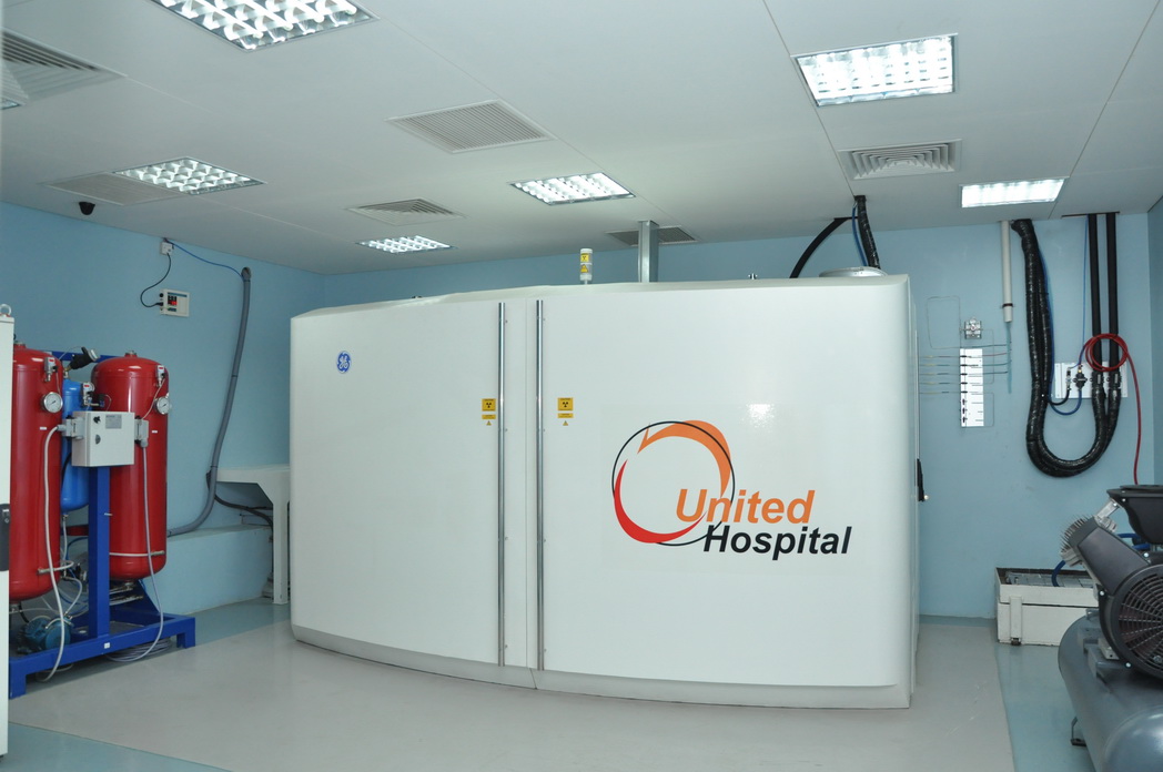
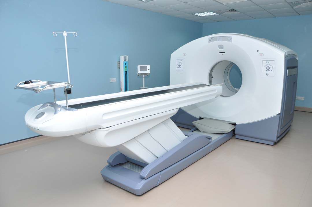
United Hospital started the first PET-CT of Bangladesh. PET (positron emission tomography) and CT (computed tomography) are both important diagnostic tools that physicians use to pinpoint disease states in the body for higher management.

PET  is a nuclear medicine and functional imaging technique that produces a three-dimensional image of functional processes in the body. The PET scan demonstrates the biological function of the body's tissues while the CT scan provides information about the body's anatomy such as size, shape, and location. By combining these two technologies, physicians can more accurately diagnose and monitor diseases such as cancer, heart diseases and certain brain disorders.

ONCOLOGY (Cancer) is the most important application of PET/CT and provides vital diagnostic information that can alter the course of cancer treatment and sometimes help in avoiding unwarranted surgery. PET can help provide critical information about whether a tumor is malignant or not, the extent of cancer, whether it has spread to other organs, monitoring of cancer recurrences and monitoring the effectiveness of treatment therapy.

CARDIOLOGY (Heart) - PET provides high level accuracy in assessing myocardial viability and perfusion.

NEUROLOGY (Brain) - PET provides highly accurate information to localize areas of the brain which cause epileptic seizures and determines if surgery is an option.

PET-CT


The advanced capabilities of PET/CT at United Hospital include:

  • Optimized system design: The highest image-sensitivity in the industry, enabling detection of disease at an earlier stage, potentially lowering the dose requirements and providing faster patient exams.
  • Motion Match: Phase-matched 4D PET and CT-fused images improve the quality of scans in areas prone to motion which can blur results such as in the lungs.
  • Patient-Friendly Design: a comfortable bed for patients up to 500 lbs., limiting the effects of claustrophobia.

In nuclear medicine imaging, radiopharmaceuticals are taken internally - intravenously or orally. Then, external detectors (gamma cameras) capture and form images from the radiation emitted by the radiopharmaceuticals. This process is unlike a diagnostic X-ray, where external radiation is passed through the body to form an image.

There are several techniques of diagnostic nuclear medicine:

•    2D: Scintigraphy ("scint") – this is the use of internal radionuclides to create two-dimensional images.
•    3D: SPECT - this is a 3D tomographic technique which uses gamma camera data from many projections and can be reconstructed in different planes.

Nuclear medicine tests differ from other imaging modes as the diagnostic tests primarily show the physiological function of the system being investigated as opposed to traditional anatomical imaging such as CT or MRI. Nuclear medicine imaging studies are generally more organ-or tissue-specific (e.g. lungs scan, heart scan, bone scan, brain scan etc.) than those in conventional radiology imaging which focus on a particular section of the body (e.g. chest X-ray, head CT scan, abdomen/pelvis CT scan etc.)

The radiopharmaceuticals used in nuclear medicine therapy emit ionizing radiation which travels only a short distance minimizing unwanted side effects and damage to non-involved organs or nearby structures. Most nuclear medicine therapies can be performed as outpatient procedures since there are few side effects from the treatment and the radiation exposure to the general public can be kept within a safe limit.