A Case of Abdominal Aortic Aneurysm
Dr. Sohely Sultana, Specialist, Radiology and Imaging Department
A 52 year old woman was admitted in the Cardiology Department of Bangabandhu Sheikh Mujib Medical University with the complaints of dysphagia for one month and exertional chest pain for two years. Her history of past illness was significant for hypertension with irregular ingestion of anti-hypertensives. She stated that A Case of Abdominal Aortic Aneurysmexcept for the last one month, she never experienced resting chest pain. She gave no H/O diabetes, severe chest pain or tearing pain in the back nor any H/O collapse or blackout.
On admission, the patient looked ill and anxious. On examination, her pulse was -92/min with radio - radial delay and decreased volume in left brachial and axillary arteries. Her blood pressure was 140/80 mm Hg in the right arm and 120/75 mm Hg in the left arm. She had normal jugular venous pressure (JVP). On auscultation her heart sounds S1+S2 were normal with no murmur. Suprasternal pulsations were present. The chest had vesicular breath sound with no added sound.
Blood investigations were unremarkable. ECG – normal, Echo Cardiography - aortic arch was dilated. X-ray chest PA view showed aneurysmal dilatation of the arch and descending aorta. USG of chest showed large mediastinal mass more remarkable in the left side which could not be separated from the aortic arch.
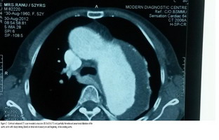
Contrast enhanced CT scan revealed massive (10.0x 8.6x 7.5 cm) partially thrombosed aneurysmal dilatation of the aortic arch with sharp kinking (bend) at the distal end of the aneurysm and the beginning of the descending aorta. Fig-1:
CT angiogram revealed a large aneurysmal dilatation of aortic arch measuring 10.0 x 8.6 x7.5 cm extending up to the beginning of the thoracic aorta with a large intramural thrombus. Wall calcifications were also noted in the dilated part. The patient was then referred to the Cardiac Surgery Department for immediate management. But the patient refused to have surgery. Two months later the patient again presented with severe agonizing chest pain and was admitted in DMCH. The patient’s condition deteriorated from then on and she did not survive.
Most people with AAA live healthy symptom free lives. The decision to undergo surgery involves weighing the risk of aneurysm rupture versus the risks of surgical procedure. While some general guidelines are suggested based upon the aneurysm size and its rate of enlargement, each treatment decision should be made as per individual case. Patients should discuss the individual risk of surgery with an experienced health care provider for a suitable remedy.