Congenital Constriction Band Syndrome: A Rare Case Report
 Dr Umme Iffat Siddiqua, Prof Dr Shahidul Islam, Dr Jan Mohammad, Dr Qamrul Hasan
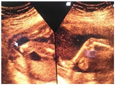
A 29 year old South African female primi gravida with 21+ weeks of pregnancy came to United Hospital for anomaly scan. This was a planned pregnancy and till this time it was uneventful. She also had no history of teratogenic drug intake or positive family history of any congenital anomaly. During USG scanning all the parameters were found to be corresponding to her LMP and amniotic fluid was also normal. But during search for anomaly it was found that the left leg of fetus just few inches below the knee was completely amputated. The remaining portion of tibia and fibula of fetus was normal in thickness though tapered down to end at amputated site.
Congenital constriction band (also known as ABS or amniotic band syndrome, amniotic band sequence, pseudoainhum, ADAM complex, amniotic band constriction, Streeter dysplasia, annular defects) is a congenital disorder caused by entrapment of fetal parts (usually a limb or digit) in fibrous amniotic bands while in utero. It may present as constriction rings around the digits, arms and legs, swelling of the extremities distal to the point of constriction (congenital lymphedema), amputation of digits, arms and legs (congenital amputation). Etiology of this syndrome is unknown; however there are two main theories. The amniotic band theory is that ABS occurs due to partial rupture of the amniotic sac. This rupture involves only the amnion; the chorion remains intact. Fibrous bands of the ruptured amnion float in the amniotic fluid and can encircle and trap some parts of the fetus. Later the fetus grows but the bands do not, rather become constricting. This constriction reduces blood circulation, hence causes congenital abnormalities and in some cases a complete natural amputation may occur. The vascular disruption theory says because the constricting mechanism does not explain the high incidence of cleft palate and other forms of cleft defects occurring together with ABS, this co-occurrence suggests an intrinsic defect of the blood circulation. Diagnosis can be made by 3D ultrasound and MRI. ABS is not genetic i.e. not inherited; so it is extremely unlikely to affect a future pregnancy. The incidence is 1 in 1200 live births. Upto 50% of cases have other congenital anomalies including club foot deformity, cleft lip and cleft palate. Hand and finger anomalies occur in upto 80% of cases. After birth of the baby, plastic and reconstructive surgery can be considered; prosthetics also may help some to live a more functional life with long term physical and occupational therapy. In rare cases, fetal surgery might be considered provided neither vital organs nor the umbilical cord of fetus were affected.